



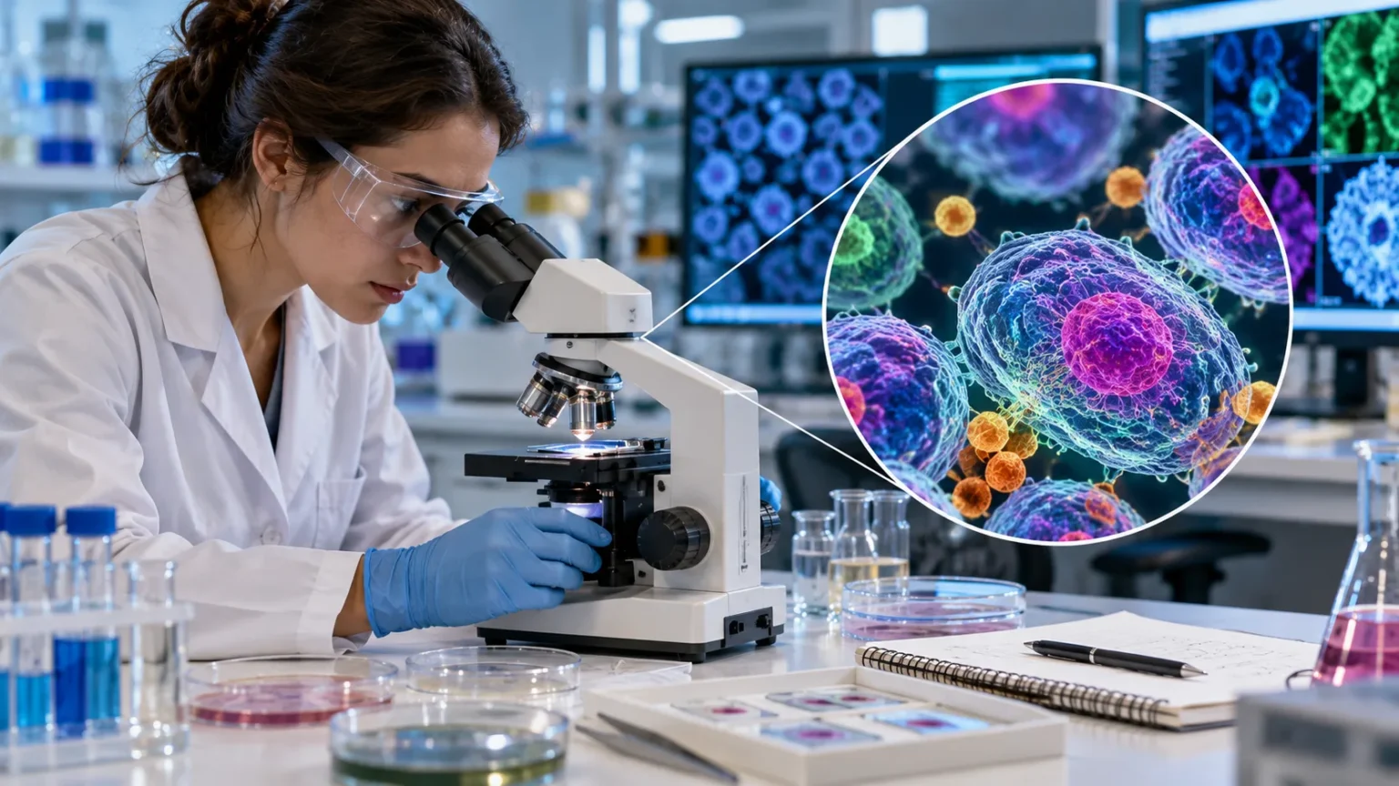
The Fundamental Principles Behind Optical Microscopes
Microscopes serve as essential tools for viewing tiny objects that are invisible to the naked human eye. Researchers rely heavily on these optical devices to study biological samples and materials in fine detail. Light passes directly through the transparent specimen to form an enlarged image for the observer. This fundamental process reveals structures that would otherwise remain hidden from view.
The history of microscopy spans several centuries of scientific innovation and engineering progress. Early inventors ground small glass beads to bend light rays and magnify small organisms. Modern instruments combine multiple glass elements to achieve much higher levels of clarity and magnification. These upgrades allow modern laboratories to push the boundaries of visual science.
Understanding how light interacts with matter forms the foundation of all optical microscopy work. Photons strike the sample and change direction depending on the density of the material. Specialized objective lenses capture these altered light rays before they reach your eyes. Every component works in harmony to produce a clear and faithful visual representation.
How Light Bends Through Glass Lenses
Light refraction occurs whenever photons pass from one medium into another of different density. Curved glass lenses exploit this physical phenomenon to bend light rays toward a single focal point. The curvature of the glass determines how strongly the light bends. Manufacturers shape these glass surfaces with extreme precision to minimize optical distortion.
Curved lenses act as the primary engine for image enlargement inside standard compound microscopes. Light rays enter the objective lens after passing through the physical slide. The lens bends these rays outward so they project a larger image further up the tube. Your eyes then view this expanded projection through the ocular lens.
Glass quality plays a massive role in determining the final sharpness of the observed image. Low-grade glass often introduces color fringes that obscure fine details on the slide. Apochromatic lenses correct these color errors by aligning different wavelengths of light perfectly. High-end optics ensure that scientists see accurate colors and sharp outlines every time.
The Vital Role of Magnification and Resolution
Magnification simply means making a small object appear much larger than its actual physical size. High magnification numbers sound impressive during public discussions about scientific equipment capabilities. However raw magnification alone cannot reveal fine structures if the image lacks sharpness. True clarity depends heavily on another optical metric known as resolution.
Resolution defines the ability of a microscope to separate two close points as distinct entities. Two small dots merge into a blurry single blob if resolution falls below required thresholds. Wavelength limits of visible light restrict how close two points can be resolved. Physics dictates that light microscopes cannot resolve details smaller than half the wavelength of light.
Numerical aperture is a crucial specification stamped onto every quality objective lens manufactured today. This value measures the ability of the lens to gather light and resolve fine sample details. Immersion oil helps increase numerical aperture by bridging the gap between slide and glass. Scientists must balance magnification and resolution to capture truly meaningful scientific data.
Exploring the Internal Parts of a Compound Microscope
A compound microscope utilizes two separate lens systems working together to enlarge specimens. The ocular lens sits at the top where the observer places their eyes. Below that component lies a rotating nosepiece holding multiple objective lenses of varying power. Each part contributes to the overall magnification capacity of the entire mechanical assembly.
The stage provides a flat stable platform where glass specimen slides rest securely during observation. Mechanical clips hold the glass slide firmly in place while knobs move it around. Beneath the stage sits a condenser lens that focuses illumination onto the sample area. An adjustable iris diaphragm controls the exact amount of light passing upward.
Focus knobs allow users to adjust the distance between the specimen and the lens. Coarse adjustment handles large movements to bring the sample into rough initial view quickly. Fine adjustment knobs make tiny movements for achieving sharp image focus on delicate structures. A sturdy metal frame supports all these optical components and keeps them properly aligned.
Venturing Beyond Light with Electron Microscopes
Standard light microscopes face physical limits imposed by the wavelength of visible light photons. Researchers needed tools that could peer deeper into the microscopic world of atoms. Electron microscopes solve this problem by replacing light beams with focused beams of electrons. Electrons possess much shorter wavelengths than visible light photons to overcome optical limits.
Magnetic fields replace glass lenses inside modern electron microscopes to guide the particle beams. Electromagnetic coils bend the fast-moving electrons with extreme precision inside a heavy vacuum column. Air molecules would scatter the electron beam so the entire tube must remain evacuated. This complex engineering setup allows researchers to achieve millions of times magnification.
Images produced by electron microscopes appear in shades of gray on computer monitors. Computers process the signals generated by electron interactions to build detailed digital representations. False colors are often added later to highlight specific biological structures for publication. These advanced devices opened entirely new frontiers in microbiology and materials science research.
Scanning Versus Transmission Electron Microscopy
Scanning electron microscopes create stunning three-dimensional images of solid specimen surfaces. A focused electron beam sweeps back and forth across the coated sample in a grid pattern. Secondary electrons bounce off the surface topography and get collected by specialized detectors. The resulting pictures show external textures with incredible depth of field and sharpness.
Transmission electron microscopes operate differently by shooting electrons straight through ultra-thin slices of material. Denser regions block more electrons while thinner areas allow particles to pass through easily. This differential transmission forms a flat two-dimensional shadow image on a fluorescent screen. Scientists use this method to study internal cellular organelles and viral structures.
Sample preparation differs significantly between scanning and transmission electron microscopy methods. Scanning samples require conductive metallic coatings to prevent electric charge buildup on surfaces. Transmission samples demand extremely thin slicing using diamond knives on specialized equipment. Both techniques require rigorous preparation steps because electron beams operate inside hard vacuums.
Peering Into the Cellular Level of Life
Cell biology relies heavily on microscopes to understand the basic building blocks of living organisms. Robert Hooke first observed cell walls in cork tissue using an early compound setup. Modern researchers can watch living cells divide and move in real time using fluorescence. These observations reveal how biological systems function at the microscopic level daily.
Staining techniques help scientists differentiate between transparent cellular components inside tissue samples. Chemical dyes bind to specific molecules like proteins or nucleic acids within the cell. Methylene blue and crystal violet make nuclei and cell membranes stand out clearly. Without these stains most biological specimens appear as featureless transparent shapes under light.
Fluorescence microscopy takes cellular observation a step further by using specific light wavelengths. Tagged antibodies attach to target proteins inside the cell and glow when illuminated. This specific labeling lets researchers track single molecules moving around living tissue. Medical researchers use this technology to identify abnormal cellular behavior in human diseases.
Observing Bacteria and Viruses in Action
Bacteria are single-celled microorganisms that researchers can view easily with standard high-power light microscopes. Oil immersion lenses bring individual bacterial cells into sharp focus for clinical identification. Microbiologists study bacterial shapes and arrangements to diagnose infections in medical laboratories. Observing these tiny organisms helps public health officials track disease outbreaks worldwide.
Viruses are much smaller than bacteria and remain invisible under traditional light microscopes. These infectious agents require powerful electron microscopes to resolve their structural protein coats. Scientists study viral morphology to understand how pathogens infect host cells and spread. This visual data aids pharmaceutical companies in designing effective vaccines and antiviral drugs.
Live imaging of bacterial motility shows how microbes swim through liquid environments using flagella. Time-lapse photography records these movements over extended periods for detailed behavioral analysis. Researchers monitor how bacteria form biofilms on medical implants and household surfaces. Understanding these survival mechanisms leads to better cleaning products and clinical treatments.
Unlocking the Secrets of Plant and Animal Tissues
Histology is the branch of biology focused on studying microscopic plant and animal tissue structures. Pathologists examine human tissue biopsies under light microscopes to detect cancerous growths. Plant scientists analyze leaf cross-sections to understand how vascular bundles transport water. Each tissue type displays unique architectural patterns that reflect its biological role.
Embedding soft tissues in paraffin wax allows technicians to slice extremely thin sections. Microtome blades cut tissue ribbons measuring only a few microns in thickness uniformly. These delicate slices are mounted on glass slides before staining and cover-slipping. This preparation preserves cellular architecture for long-term storage and clinical review.
Animal organ systems show remarkable organization when viewed through specialized microscopy equipment. Muscle tissue reveals repeating striped patterns that explain how contraction generates physical force. Neural tissue displays branching networks of neurons transmitting electrical signals through the body. These structural insights connect microscopic anatomy directly to macroscopic physiological functions.
Examining Materials and Nanostructures
Material science uses advanced microscopes to inspect metals and polymers at microscopic scales. Metallurgists check crystal grain structures in steel beams to ensure structural safety. Surface defects and micro-cracks become visible before catastrophic failures happen in industrial settings. Quality control departments rely on these inspections to maintain high manufacturing standards.
Semiconductor manufacturing requires extreme precision that only specialized microscopy tools can provide safely. Engineers check silicon wafer surfaces for microscopic dust particles during computer chip production. Atomic force microscopes drag tiny physical probes across surfaces to map atomic contours. These tools ensure that nanoscale electronic circuits function correctly without short circuits.
Nanotechnology creates engineered materials with unique properties derived from their microscopic architecture. Carbon nanotubes and graphene sheets require electron microscopy to verify their structural integrity. Researchers study nanoparticle delivery systems for targeted medicine using high-resolution imaging. Visual verification remains essential for advancing modern engineering and materials science.
Preparing Samples for Clear Observation
Sample preparation is often the most critical step in successful microscopy work. Poorly prepared slides yield blurry images regardless of how expensive the microscope lens is. Technicians must clean glass slides thoroughly before placing any biological specimen on top. Correct mounting ensures that light passes through the sample without unwanted scattering.
Fixation chemicals lock cellular structures in place before decay alters the natural tissue state. Formaldehyde solutions cross-link proteins to preserve internal components in a lifelike condition. Dehydration steps remove water from samples using graded alcohol baths carefully. These preservation methods protect fragile biological specimens during subsequent staining procedures.
Cover slips seal the mounted specimen against air exposure and physical damage. Clear mounting media matches the refractive index of glass to optimize light transmission. Proper sealing prevents bubble formation that ruins optical clarity during observation sessions. Meticulous preparation habits separate amateur attempts from professional scientific imaging results.
Conclusion
Microscopes have transformed human understanding of the natural world significantly over the past centuries. From simple glass beads to advanced electron scanners these instruments bridge the gap between invisible realms and human perception. Scientists continue pushing optical boundaries to uncover deeper secrets hidden within cells and materials.
Understanding how these optical devices function helps students and researchers appreciate the complexity of science. Proper handling sample preparation and optical alignment remain fundamental skills across all laboratory settings. Investment in high-quality microscopy equipment directly drives medical breakthroughs and technological innovations worldwide.
The future of microscopy promises even greater resolution and non-invasive imaging capabilities for researchers. As digital technology and optical engineering merge new possibilities emerge for exploring life and matter. Curiosity combined with powerful magnification tools will keep expanding human knowledge for generations ahead.
Frequently Asked Questions
How do light microscopes work?
Light microscopes use glass lenses to bend and focus visible light passing through a transparent specimen. This creates an enlarged, clear image for the observer.
What is the difference between magnification and resolution?
Magnification makes an object look larger on screen or through optical lenses. Resolution defines the fundamental ability to separate two close points distinctly as individual entities.
Can light microscopes see viruses?
Standard light microscopes cannot see individual viruses due to the physical wavelength limits of visible light. Researchers must use powerful electron microscopes to view viral structures.
Why is oil used in microscopy?
Immersion oil bridges the gap between the glass slide and the objective lens. This prevents light refraction loss and increases numerical aperture for sharper high-power focus.
How do electron microscopes achieve higher resolution?
Electron microscopes use focused beams of high-speed electrons instead of standard light photons. Electrons possess much shorter wavelengths, which overcomes the optical limits of visible light.