



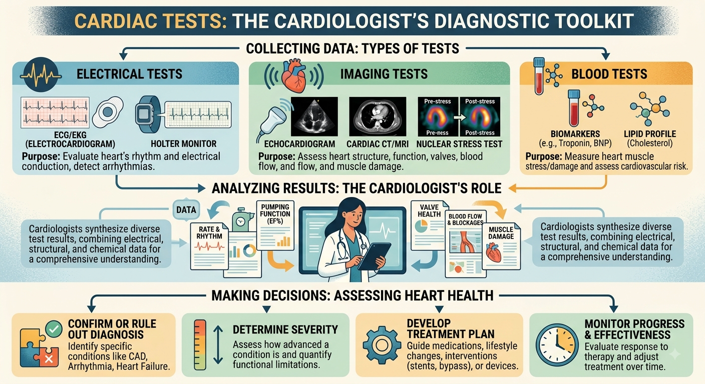
Cardiac tests help doctors assess different aspects of the heart, including its electrical activity, rhythm, structure, pumping function, blood flow and coronary arteries. Patients may be advised to undergo cardiac testing because of symptoms such as chest pain, shortness of breath, palpitations, dizziness or reduced exercise tolerance, or because they have cardiovascular risk factors or abnormal findings from an earlier examination.
The type of test recommended depends on the symptoms, medical history, cardiovascular risk profile and clinical question. Some tests assess the heart while a patient is resting, while others examine what happens during physical exertion or provide images of the heart and its blood vessels.
Understanding the purpose of common cardiac tests can help patients know what information a cardiologist may be looking for during a heart assessment.
What Does a Cardiologist Assess?
A cardiologist is a doctor who assesses, diagnoses and manages conditions affecting the heart and cardiovascular system.
During a consultation, the cardiologist may ask about symptoms, medical history, medication, family history and cardiovascular risk factors. A physical examination may also be performed before deciding whether cardiac testing is appropriate.
Tests may be used to assess:
- Heart rate and rhythm
- Electrical activity of the heart
- Heart chamber size
- Heart pumping function
- Heart valves
- Heart-muscle movement
- Blood pressure
- Blood flow to the heart
- Coronary artery narrowing
- Exercise response
- Cardiovascular risk factors
A single test does not examine every part of heart health. Several investigations may therefore be considered when different types of information are needed.
1. Electrocardiogram
An electrocardiogram, commonly called an ECG or EKG, records the electrical activity of the heart.
Small electrodes are attached to the chest and limbs to detect electrical signals produced with each heartbeat. These signals are displayed as a tracing that a doctor can review.
An ECG may provide information about:
- Heart rate
- Heart rhythm
- Certain arrhythmias
- Electrical conduction abnormalities
- Changes that may occur with heart-muscle injury
- Certain signs of strain affecting the heart
A doctor may recommend an ECG for symptoms such as:
- Chest pain
- Palpitations
- Dizziness
- Fainting
- Shortness of breath
- Irregular heartbeat
An ECG records the heart during a relatively short period. If symptoms occur intermittently, another form of rhythm monitoring may be needed.
2. Echocardiogram
An echocardiogram uses ultrasound waves to produce moving images of the heart.
A cardiologist may recommend this test when information about the heart’s structure or function is required.
An echocardiogram may assess:
- Heart chamber size
- Heart-wall thickness
- Pumping function
- Heart-muscle movement
- Heart valve structure
- Valve narrowing or leakage
- Blood flow through the heart
- Fluid around the heart
- Selected structural heart conditions
The test may be considered for patients with:
- Shortness of breath
- Heart murmur
- Suspected valve disease
- Heart failure symptoms
- Cardiomyopathy
- Abnormal findings from another heart test
- Certain changes after a heart attack
A standard echocardiogram does not directly show every narrowing within the coronary arteries. Other investigations may be required when coronary artery disease is suspected.
3. Holter and Ambulatory ECG Monitoring
Palpitations and abnormal heart rhythms do not always occur during a routine consultation.
Holter monitoring records the heart’s electrical activity continuously over a longer period than a standard ECG. The patient wears a portable recording device while continuing with usual daily activities.
It may be used to investigate:
- Intermittent palpitations
- Irregular heartbeat
- Unexplained dizziness
- Fainting
- Episodes of rapid heartbeat
- Suspected rhythm disturbances
Patients may be asked to record when symptoms occur so that the timing can be compared with the ECG recording.
Other ambulatory rhythm monitors may be used for different lengths of time depending on how frequently symptoms occur.
4. Cardiac Stress Test
A cardiac stress test examines how the heart responds when its workload increases.
Exercise testing is commonly performed using a treadmill or stationary bicycle. Heart rhythm, blood pressure, ECG changes and symptoms can be monitored while exercise intensity gradually increases.
A stress test may be considered for:
- Chest discomfort during exertion
- Breathlessness during exercise
- Reduced exercise tolerance
- Exercise-related palpitations
- Assessment of suspected coronary artery disease
- Exercise assessment in selected patients with existing heart conditions
Different forms of stress testing may be used.
Exercise ECG
An exercise ECG records the heart’s electrical activity during physical exertion.
Stress Echocardiogram
A stress echocardiogram combines increased cardiac workload with ultrasound imaging to assess how the heart muscle moves and functions under stress.
Nuclear Stress Test
Nuclear stress testing combines stress with cardiac imaging to assess blood flow to the heart muscle.
Patients who cannot exercise adequately may sometimes undergo stress testing using medication instead of physical exercise.
The type of stress test depends on the patient’s health and the clinical information required.
5. Ambulatory Blood Pressure Monitoring
A blood pressure reading taken during a consultation shows the patient’s blood pressure at that particular time.
Ambulatory blood pressure monitoring records readings repeatedly while the patient continues with normal daily activities and during sleep.
It may help assess:
- Blood pressure throughout the day
- Night-time blood pressure
- Variation in blood pressure
- Readings outside the clinic environment
- Response to blood pressure treatment in selected cases
High blood pressure can place strain on the cardiovascular system over time.
Blood pressure results are usually interpreted alongside other factors such as age, cholesterol, diabetes, smoking history, family history, and existing cardiovascular disease.
6. Blood Tests
Blood tests can provide information about cardiovascular risk factors and medical conditions that may affect the heart.
Depending on the clinical situation, a cardiologist may review:
- Cholesterol levels
- Blood glucose
- Kidney function
- Electrolytes
- Full blood count
- Selected cardiac biomarkers
- Thyroid function in relevant cases
For example, cholesterol and blood glucose results may form part of cardiovascular risk assessment, while certain cardiac biomarkers may be measured when heart-muscle injury is suspected.
Blood tests do not replace cardiac imaging or electrical tests. They provide another part of the information used during cardiovascular assessment.
7. CT Coronary Calcium Score
A coronary calcium scan is a type of CT scan that measures calcium within the coronary arteries.
Calcium can be associated with atherosclerotic plaque within these arteries.
The test may be considered as part of cardiovascular risk assessment in selected individuals.
A coronary calcium score provides information about calcified coronary plaque but does not provide the same information as CT coronary angiography or invasive coronary angiography.
The decision to use this test depends on the patient’s cardiovascular risk profile and whether the result is expected to assist clinical decision-making.
8. CT Coronary Angiography
CT coronary angiography uses computed tomography and contrast material to produce images of the coronary arteries.
The coronary arteries supply blood to the heart muscle.
This type of cardiac imaging may help assess:
- Coronary artery anatomy
- Atherosclerotic plaque
- Narrowing within the coronary arteries
- Selected cardiac and vascular structures
CT coronary angiography may be considered when coronary artery disease is suspected and anatomical information about the coronary arteries is needed.
It differs from a cardiac stress test because the tests answer different questions.
A stress test examines the heart’s response to increased workload, while CT coronary angiography examines the anatomy of the coronary arteries.
9. Cardiac MRI
Cardiac magnetic resonance imaging, or cardiac MRI, uses magnetic fields and radio waves to produce images of the heart and surrounding structures.
Depending on the clinical indication, it may provide information about:
- Heart chamber size
- Pumping function
- Heart-muscle thickness
- Heart-muscle movement
- Areas of cardiac scarring
- Certain cardiomyopathies
- Selected structural heart conditions
- Inflammation affecting the heart muscle
Cardiac MRI does not use ionising radiation.
It may be recommended when another investigation identifies a finding that requires further characterisation or when specific information about heart muscle or structure is needed.
Not every patient undergoing cardiovascular assessment requires cardiac MRI.
10. Nuclear Cardiac Imaging
Nuclear cardiac imaging uses a small amount of radioactive tracer together with specialised imaging equipment to assess selected aspects of heart function or blood flow.
Tests may include myocardial perfusion imaging and cardiac PET.
Myocardial Perfusion Imaging
This test assesses blood flow to the heart muscle.
Images may be taken at rest and during cardiac stress to compare how blood reaches different parts of the heart muscle.
Cardiac PET
Cardiac positron emission tomography may be used in selected circumstances to evaluate heart-muscle blood flow, metabolism or tissue characteristics.
Nuclear cardiac imaging is not routinely required for every patient. Its use depends on the cardiovascular condition being investigated and the information required.
11. Coronary Angiography
Coronary angiography is an invasive procedure used to visualise the coronary arteries.
A thin catheter is introduced through a blood vessel, commonly through the wrist or groin, and guided towards the coronary arteries. Contrast material is then injected while X-ray images are taken.
Coronary angiography can provide information about:
- Coronary artery anatomy
- Location of arterial narrowing
- Severity of narrowing
- Number of coronary arteries affected
- Blood flow through the coronary arteries
The procedure may be recommended when direct assessment of the coronary arteries is clinically required.
If significant coronary narrowing is identified, the findings may guide discussion about medication, angioplasty, stent placement or referral for another form of treatment depending on the patient’s condition.
How Do Cardiologists Decide Which Test to Recommend?
The recommended cardiac test depends on the patient’s symptoms and cardiovascular risk factors.
Factors that may influence the decision include:
- Type of symptoms
- When symptoms occur
- Duration of symptoms
- Medical history
- Family history of cardiovascular disease
- Blood pressure
- Cholesterol
- Diabetes
- Smoking history
- Existing cardiovascular conditions
- Previous ECG findings
- Previous imaging results
- Ability to exercise
- Kidney function when contrast material is being considered
For example, palpitations may initially be assessed with an ECG or Holter monitor. A heart murmur may lead to an echocardiogram, while symptoms suggesting coronary artery disease may require stress testing, cardiac imaging, or coronary angiography depending on the individual clinical situation.
Patients can ask:
- Why is this test being recommended?
- What part of the heart does it assess?
- What can the test show?
- What are its limitations?
- Do I need to prepare beforehand?
- How will the result affect my care?
- Could another test be needed afterwards?
Understanding the purpose of an investigation can help patients follow the assessment process.
When Should You See a Cardiologist?
You may consider a cardiology assessment if you have:
- Chest pain or chest discomfort
- Shortness of breath
- Palpitations
- Dizziness or fainting
- Reduced exercise tolerance
- Leg swelling
- High blood pressure
- High cholesterol alongside cardiovascular risk factors
- An abnormal ECG
- An abnormal heart-screening result
- A heart murmur
- A known arrhythmia
- Known coronary artery disease
- Previous heart attack
- Existing heart failure
- Known valve disease
People searching for the best cardiologist in Singapore may be trying to identify a doctor who can investigate a particular symptom or cardiovascular concern. Rather than relying on a ranking, factors such as specialist registration, relevant clinical scope, available cardiac investigations, and arrangements for follow-up can be considered.
Urgent medical care is required for symptoms such as severe or persistent chest pain, sudden significant shortness of breath, sudden collapse, or other symptoms suggesting an acute cardiovascular emergency.
How Cardiac Tests Work Together
Different cardiac tests provide different types of information.
For example:
- An ECG assesses electrical activity.
- A Holter monitor records heart rhythm over a longer period.
- An echocardiogram assesses heart structure and function.
- A stress test examines the response to increased workload.
- CT coronary angiography assesses coronary artery anatomy.
- Cardiac MRI provides information about heart structure, function and selected tissue characteristics.
- Nuclear imaging can assess heart-muscle blood flow or function.
- Coronary angiography directly visualises the coronary arteries.
One test may therefore be followed by another if additional information is needed.
A normal result also needs to be interpreted according to what that particular test assesses. For example, a normal resting ECG does not examine heart valves or directly visualise the coronary arteries.
Cardiac tests help cardiologists assess different aspects of heart health, from electrical rhythm and blood pressure to heart structure, pumping function, and coronary blood flow.
The appropriate test depends on symptoms, medical history, cardiovascular risk factors, examination findings, and previous results. Some patients may require a single investigation, while others may undergo several tests to answer different clinical questions.
Frequently Asked Questions
What cardiac tests may a cardiologist recommend?
A cardiologist may recommend an ECG, echocardiogram, Holter monitoring, cardiac stress testing, blood pressure monitoring, blood tests, CT coronary imaging, cardiac MRI, nuclear cardiac imaging, or coronary angiography depending on symptoms and risk factors.
What is the difference between an ECG and an echocardiogram?
An ECG records the electrical activity of the heart. An echocardiogram uses ultrasound to produce images of the heart’s structure, valves, and function.
What cardiac test may be used for chest pain?
The test depends on the clinical situation. Assessment may involve an ECG, blood tests, echocardiography, stress testing, CT coronary angiography, or coronary angiography.
Can an echocardiogram detect blocked coronary arteries?
A standard echocardiogram does not directly visualise every coronary artery narrowing. Other investigations may be considered when coronary artery disease is suspected.
What is the difference between CT coronary angiography and coronary angiography?
CT coronary angiography uses a CT scanner and contrast material to produce images of the coronary arteries. Invasive coronary angiography uses a catheter inserted into a blood vessel together with contrast and X-ray imaging.
Why might a cardiologist recommend several heart tests?
Different tests assess different aspects of cardiovascular health. One test may examine rhythm while another examines heart structure, exercise response, or coronary artery anatomy.
This article is for general information only and should not replace medical advice from a qualified healthcare professional.